Research area
Molecular Docking & Drug Discovery
Questions we can help you answer
- Is docking appropriate for my question?
- Which protein structure should I use?
- How do I validate a docking protocol?
- How should network-pharmacology results be filtered and reported?
Methods we advise on
- Receptor and ligand preparation
- Re-docking validation
- Interaction analysis
- Protein–protein interaction networks
- GO/KEGG enrichment
- MD planning
Please note
- Docking scores are not binding affinities, and in silico results are not evidence of efficacy or safety.
From mechanism to structure
See the science
A signalling pathway explained step by step, and the real experimental structure of a drug bound to its target.
Mechanism · animated schematic
How a growth-factor receptor switches on — and how a kinase inhibitor stops it
- 1A growth factor binds the extracellular domain of each receptor.
- 2Ligand-bound receptors pair up (dimerise) in the membrane.
- 3The two kinase domains phosphorylate tyrosines on each other (trans-autophosphorylation).
- 4Phosphotyrosines recruit adaptor proteins (Grb2·SOS), which activate RAS.
- 5RAS → RAF → MEK → ERK relays the signal to the nucleus, switching on genes for proliferation and survival.
Simplified schematic, not to scale; many more proteins take part. Based on Lemmon MA, Schlessinger J. Cell signaling by receptor tyrosine kinases. Cell 2010;141(7):1117–1134. doi:10.1016/j.cell.2010.06.011
Interactive · loads about 210 KB · PDB 1M17
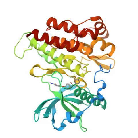
Experimental structure · PDB 1M17
Erlotinib in the EGFR kinase domain
Erlotinib, an approved EGFR inhibitor, bound in the ATP pocket of the EGFR kinase domain. Its quinazoline ring sits against the hinge region, where the backbone of Met769 lies within hydrogen-bonding distance.
Measured contacts (≤ 3.5 Å, N/O atoms)
- Met769 N — erlotinib N2: 2.7 Å
- Pro770 O — erlotinib O4: 3.21 Å
- Gly772 N — erlotinib O4: 3.24 Å
- Leu694 O — erlotinib O2: 3.27 Å
Residues within 4.5 Å of the ligand
Leu694 · Val702 · Ala719 · Lys721 · Glu738 · Leu764 · Ile765 · Thr766 · Gln767 · Leu768 · Met769 · Pro770 · Phe771 · Gly772 · Leu820 · Thr830 · Asp831
X-ray diffraction, 2.6 Å resolution. Source: RCSB PDB 1M17 — Stamos, J., Sliwkowski, M.X., Eigenbrot, C. Structure of the epidermal growth factor receptor kinase domain alone and in complex with a 4-anilinoquinazoline inhibitor. J.Biol.Chem. 2002. doi:10.1074/jbc.M207135200
Distances were measured by Rehbar from the deposited coordinates (prepared 2026-10-16). They indicate likely hydrogen bonds; they are not an energy calculation, and this is published structural data — not Rehbar’s own experiment or a client result.
Relevant services
Computational Biology & Bioinformatics
Docking, network pharmacology and reproducible analysis workflows — with honest limits on what in silico results mean.
- For:
- MPhil and PhD researchers adding an in silico chapter
- Lead:
- Scientific Lead

Scientific & Research Consulting
Sharpen the research question, choose a defensible design and plan the work before time and reagents are spent.
- For:
- Postgraduate researchers and early-career scientists
- Lead:
- Scientific Lead
Tell us what you’re working on.
Send a short, confidential summary. We reply with an initial scope assessment, and you agree deliverables, timeline and price before anything starts.